Peak Proteins Signs Agreement with University of Leeds to Access Cryo-EM Facilities.
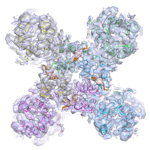
Peak Proteins is now in a position where, in collaboration with the University of Leeds, we would be able to talk about cryo-EM approaches with clients having entered into an agreement with the University to access their state-of-the-art cryo-EM facilities and expertise. This allows us to offer our clients the possibility of obtaining structural information on a range of protein targets that do not crystallise, for example large membrane proteins and protein assemblies.
Cryogenic electron microscopy (cryo-EM) is an electron microscopy (EM) technique applied on samples cooled to cryogenic temperatures and embedded in an aqueous environment. An aqueous sample solution is applied to a grid-mesh and plunge-frozen in liquid ethane.
The field of cryo-EM has developed rapidly in the last several years. Numerous technical advances, including the introduction of direct electron detectors coupled to the development of algorithms that correct for beam-induced movement and specimen drift, have enabled the wide adoption of the technique for many structural biology applications. In many instances, cryo-EM is now the first go-to method for structural analysis of certain biological samples.
In 2017, the Nobel Prize in Chemistry was awarded to Jacques Dubochet, Joachim Frank, and Richard Henderson “for developing cryo-electron microscopy for the high-resolution structure determination of biomolecules in solution.”

